43 fluorescent labels and light microscopy
Novel Fluorescent Label Shines a Light on DNA Structure in ... Liu and her team formulated a new label called Hoechst-Cy5 to overcome the hurdle that fluorescent dyes didn't work well on DNA or in processed clinical cancer samples. After showing that the new... Label-free prediction of three-dimensional fluorescence ... Fluorescence microscopy can resolve subcellular structure in living cells, but is expensive, slow, and toxic. Here, we present a label-free method for predicting 3D fluorescence directly from transmitted light images and demonstrate its use to generate multi-structure, integrated images.
Fluorescence Imaging - Teledyne Photometrics Fluorescent molecules (known as fluorophores) are used to label samples, and fluorophores are available that emit light in virtually any color. In a fluorescent microscope, a sample is labeled with a fluorophore, and then a bright light ( excitation light) is used to illuminate the sample, which gives off fluorescence ( emission light ).
Fluorescent labels and light microscopy
Label-free prediction of three-dimensional fluorescence ... Although fluorescence microscopy can resolve subcellular structure in living cells, it is expensive, is slow, and can damage cells. We present a label-free method for predicting three-dimensional fluorescence directly from transmitted-light images and demonstrate that it can be used to generate multi-structure, integrated images. A quick guide to light microscopy in cell biology - PMC Fluorescence microscopy uses fluorescent dyes (fluorophores), which are molecules that absorb one wavelength of light (the excitation wavelength) and emit a second, longer wavelength of light (the emission wavelength). Different Ways to Add Fluorescent Labels | Thermo Fisher ... Using fluorescence provides greater contrast compared to viewing your samples with brightfield microscopy alone. Labeling various targets with separate fluorescent colors allows you to visualize different structures or proteins within a cell in the same experiment.
Fluorescent labels and light microscopy. Fluorescent Labelling - an overview | ScienceDirect Topics Fluorescence microscopy Fluorescent labeling methods are generally based on reactive derivatives of fluorophores that selectively bind to functional groups contained in target biomolecules and are widely used in biotechnology because of their non-destructive properties and the high sensitivity of fluorescence techniques ( Sahoo, 2012 ). Label-free prediction of three-dimensional fluorescence ... We present a label-free method for predicting three-dimensional fluorescence directly from transmitted-light images and demonstrate that it can be used to generate multi-structure, integrated... Fluorescent Labeling - What You Should Know - PromoCell Fluorescence microscopy allows the identification of cells and cellular components and the monitoring of cell physiology with high specificity. Fluorescence microscopy separates emitted light from excitation light using optical filters. The use of two indicators also allows the simultaneous observation of different biomolecules at the same time. Fluorescent Dyes | Science Lab | Leica Microsystems A basic principle in fluorescence microscopy is the highly specific visualization of cellular components with the help of a fluorescent agent. This can be a fluorescent protein - for example GFP - genetically linked to the protein of interest. If cloning is impossible - for instance in histologic samples - techniques such as immunofluorescence staining are used to visualize the protein ...
Fluorescence Microscope: Principle, Types, Applications ... Fluorescence microscopy is a type of light microscope that works on the principle of fluorescence. A substance is said to be fluorescent when it absorbs the energy of invisible shorter wavelength radiation (such as UV light) and emits longer wavelength radiation of visible light (such as green or red light). Fluorescence Microscopy & Cell Imaging | Research | UNM ... The Fluorescence Microscopy and Cell Imaging Shared Resource provides UNM researchers access to state-of-the art instrumentation for multiple fluorescence and transmitted light microscopy techniques: Laser scanning single and multi-photon microscopes and hyperspectral imaging systems enable simultaneous visualization and quantification of ... Light Sheet Fluorescence Microscopy - an overview ... Applications of single-molecule fluorescence microscopy. (A) The photophysical properties of a fluorophore contain information about its position and its state. This allows, for example, tracking molecules, observing conformational and constitutional changes, or following chemical reactions. (B) Examples for applications in biology and chemistry. Researchers Develop New Fluorescent DNA Label for Clinical ... Superresolution fluorescence microscopy creates clearer biological images through the blinking on and off of specific labels at different times, allowing a composite image to be reconstructed showing all of the labeled components in high resolution.
Fluorescence microscopy: established and emerging methods ... The primary concern in all forms of microscopy is the generation of contrast; for fluorescence microscopy contrast can be thought of as the difference in intensity between the cell and background, the signal-to-noise ratio. High information-content images can be formed by enhancing the signal, suppressing the noise, or both. Label-free prediction of three-dimensional fluorescence ... Label-free prediction of three-dimensional fluorescence images from transmitted-light microscopy Understanding cells as integrated systems is central to modern biology. Although fluorescence microscopy can resolve subcellular structure in living cells, it is expensive, is slow, and can damage cells. Researchers demonstrate label-free super-resolution microscopy A newly developed sub-diffraction-limit microscopy approach doesn't require fluorescent labels. The video shows the process of the data evaluation algorithm, retrieving the positions and sizes ... What do researchers use fluorescent labels and light ... What do reseaches use fluorescent labels and light microscopy to do? A) produce movies of cells as they grow, divide and develop B) scan cells with laser beans C) follow molecules moving through ...
Dots, Probes and Proteins: Fluorescent Labels for Microscopy and Imaging
New fluorescent label provides a clearer picture of how ... A molecule of interest is labelled with a special fluorescent dye that flashes on and off like a blinking star. Unlike traditional fluorescence microscopy, which uses labels that glow constantly,...

What is Correlative Light and Electron Microscopy?
Fluorescent tag - Wikipedia Fluorescent labels can be hybridized to mRNA to help visualize interaction and activity, such as mRNA localization. An antisense strand labeled with the fluorescent probe is attached to a single mRNA strand, and can then be viewed during cell development to see the movement of mRNA within the cell. Fluorogenic labels

ImageCORE Microscopy Facility - Confocal Basics
Fluorescence microscope - Wikipedia A fluorescence microscope is an optical microscope that uses fluorescence instead of, or in addition to, scattering, reflection, and attenuation or absorption, to study the properties of organic or inorganic substances. "Fluorescence microscope" refers to any microscope that uses fluorescence to generate an image, whether it is a simple set up like an epifluorescence microscope or a more ...

Cryo-electron microscopy (EM) Work Flow. A purified sample is applied... | Download Scientific ...
Imaging Flies by Fluorescence Microscopy: Principles ... The development of fluorescent labels and powerful imaging technologies in the last two decades has revolutionized the field of fluorescence microscopy, which is now widely used in diverse scientific fields from biology to biomedical and materials science. ... has brought about the era of fluorescence light microscopy. The first fluorescence ...

JC-1 - CAS 47729-63-5 - Calbiochem A cationic, fluorescent, carbocyanine dye that can be used as ...
In Silico Labeling: Predicting Fluorescent Labels in ... Fluorescence microscopy images can be predicted from transmitted-light z stacks • 7 fluorescent labels were validated across three labs, modalities, and cell types • New labels can be predicted using minimal additional training data Summary Microscopy is a central method in life sciences.
Super-Resolution Optical Microscopy Nanoscopy And Nanopositioning Motion Control
Integrating High Resolution Light Microscopy and Real Time ... Integrating High Resolution Light Microscopy and Real Time Observation of Fluorescent Labels - Volume 14 Issue 6 Skip to main content Accessibility help We use cookies to distinguish you from other users and to provide you with a better experience on our websites.

A schematic diagram of the light path in the ptychographic microscope... | Download Scientific ...
Fluorescence Microscopy vs. Light Microscopy This means that fluorescent microscopy uses reflected rather than transmitted light. For example, a commonly used label is green fluorescent protein (GFP), which is excited with blue light and...

Antigen ab reactions
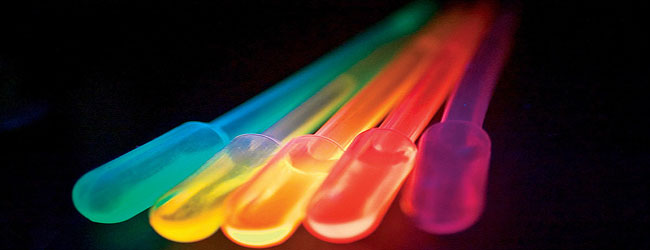
Fluorescence Microscopy Fig. 1. Fluorite, fluorescent under UV light Fluorescence is the temporary absorption of electromagnetic wavelengths from the visible light spectrum by fluorescent molecules, and the subsequent emission of light at a lower energy level. Stimulating light excites an electron, raising energy to an unstable level.
Post a Comment for "43 fluorescent labels and light microscopy"